Flash News

Lungs Anatomy

Anatomy Of Lungs

  • Lungs are a pair of conical shaped reddish grey colored organs present in the thoracic cavity on either side of the heart.
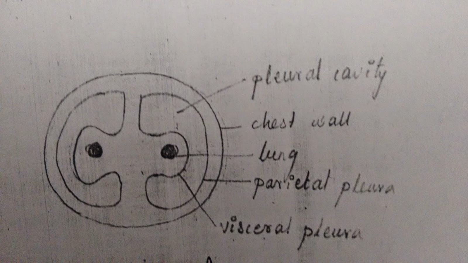
  • Each lung is covered by a serous membrane called as pleura.
  • The pleura is composed of 2 layers,
  • Outer parietal pleura and Inner visceral pleura.
  • Between them a space is present called as pleural cavity which is filled with pleural fluid.
  • Parietal pleura is divided into 4 parts,
    Costal pleura, diaphragmatic pleura, mediastenal pleura and cervical pleura.
  • At birth, these lungs are grayish pink in color, as age advances due to the deposition of carbon particles the lungs will become grayish black in color.
  • Each lung is conical in shape with a narrow apex above and a broad based below.
  • The apex extends into the lower part of the neck.
  • The base is concave and is related to the domes of the diaphragm.
  • The diaphragm separates base of left lung from stomach and spleen.
  • Each lung has territory borders, Anterior, posterior and inferior and 3 surfaces, Medial, lateral or costal and inferior.
  • The anterior border of the lung is straight where as that of left lung is deviated in the lower part called as the cardiac notch.


  • In the lower part it shows a tongue shaped projection called as lingual.
  • The posterior border is rounded.
  • The inferior surface is also called as base.
  • The lateral or the costal surfaces shows impressions caused by the ribs.
  • On the medial surface there is a depression called as hilum which is the entry and exit point of lung.
  •  Hilum of lung will give passage to bronchus, pulmonary artery and secondary pulmonary veins.
  • Blood Supply : By bronchial arteries.
  • Venous Drainage : By bronchial veins.
  • Nerve Supply : Vagous nerves.

Fissures and Lobes of Lungs :


  • Right lung has tow fissures, oblique fissure and horizontal fissure dividing it into 3 lobes upeer lobe, middle lobed and lower lobe.
  • Left lung has one fissure, oblique fissure dividing it into 3 lobes upper lobe, middle lobed and lower lobe.

Differences Between Right and Left lungs :

Right Lung :

  •         Shorter, broader and has more volume.
  • ·        Anterior border is straight.
  • ·        Has two fissures and 3 lobes.

Left Lung :

  • ·        Longer, narrower with less volume.
  • ·        Anterior border is cardiac notch.
  • ·        One fissure and 2 lobes

 Applied Anatomy :

  • Inflammation of lung is called pneumonia.
  • Inflammation of bronchi is bronchitis.
  • In a condition called as bronchial asthma the bronchi are narrowed there by causing difficulty in breathing.

 Related Posts :


Bronchial Tree Anatomy

Anatomy Of Bronchial Tree 

  • The trachea at the level of sternal angle will divide into two primary branchi.
  • Right bronchus is shorter, wider and vertical.
  • Left bronchus is longer, narrower and oblique.
  • Each Primary bronchus will divide into secondary bronchi which will supply each lobe of the lung.
  • On the right side there are 3 secondary bronchi and on left side there  are 2 secondary bronchi.
  • Each secondary bronchus will divide into teritiory bronchi which will further divide into teritiory bronchioles, alveolar ducts, alveolar sac, and finally alveoli.
  • These are the functional units of lung where exchange of gases will occur.


Trachea Anatomy

Anatomy Of Trachea

  • It starts as a continuation downwards of larynx in the lower part of front of neck at the level of C6 vertebra.
  • It passes downwards enters the thorax and at the level of sternal angle it end by dividing into two bronchi.


  • The trachea is composed of 15 to 20 ‘C’ shaped cartilaginous rings.


  • The posterior part of ring is deficient which is filled by a muscle called as tracheolis.
  • These rings are connected to each other by membranes there by forming a tube is trachea.

Microscopic Structure of Trachea :

  • The wall of the trachea is composed of 4 layers, inside outwards they are mucosa, sub mucosa, cartilage and adventitia.
  • The mucosa is lined by pseudostratified ciliated columnor epithelium with goblet cells.
  • The cartilage is composed of hyaline cartilage.

Applied Anatomy :

Tracheasotomy : In obstructions of the upper part of the respiratory system an opening is made in the trachea and a tube is inserted through which the person will be able to breathe. This procedure is called as tracheaostomy.

Larynx Anatomy

Anatomy Of Larynx

  •  It is an organ of respiration and also phonation ( speech ).
  • It is located in the upper part of the front of the neck in the midline.
  •  It starts at the level of L3 vertebra and ends at the level of C6 vertebra by being continued as trachea.
  • It is a longer in males than in females.
  • It is composed of mostly cartilages which are connected to each other by muscles, membranes, ligaments and joint.
  • Cartilages of larynx are 9 in number, 
    3 unpaired thyroid cartilage, cricoids cartilage and epiglottis.
    3 paired arystenoid cartilages, uneiform cartilages, corniculate cartilages.
  • Thyroid cartilage is largest of all.
  • Cricoid cartilage : signet ring shaped.
  • Epiglottis : Leaf Like.


  • Within the cavity of larynx, there are 2 pairs of folds, upper pair of vestiblar folds and lower pair of vocal folds.
  • The movements of vocal folds are responsible for the production of voice
  • These folds are dividing the cavity of larynx into 3 parts Supra glottis, glottis and infraglottis.
  • Arterial Supply : Superior and inferior laryngeal arteries.
  • Nerve Supply : Recurrent laryngeal nerve.

Applied Anatomy :

Aphonia : Injury to the recurrent laryngeal nerve will cause paralysis of vocal folds there by causing loss of voice. This condition is called as aphonia.

Related posts :

Urinary System

URINARY SYSTEM

     This system is composed of a pair of kidneys, where urine is formed, a pair of ureters through which the urine formed in the kidneys will pass downwards, an urinay bladder where the urine is stored and an urethra through which the urine is excreted out.

Kidney Anatomy

Nephron Anatomy.

Ureter Anatomy 

Urethra Anatomy

Urinary Bladder Anatomy

Related Posts :